파일:Histology bse.jpg
누리위키, 온 누리의 백과사전
최대 해상도입니다.
Histology_bse.jpg (700 × 558 픽셀, 파일 크기: 73 KB, MIME 종류: image/jpeg)
| 설명 |
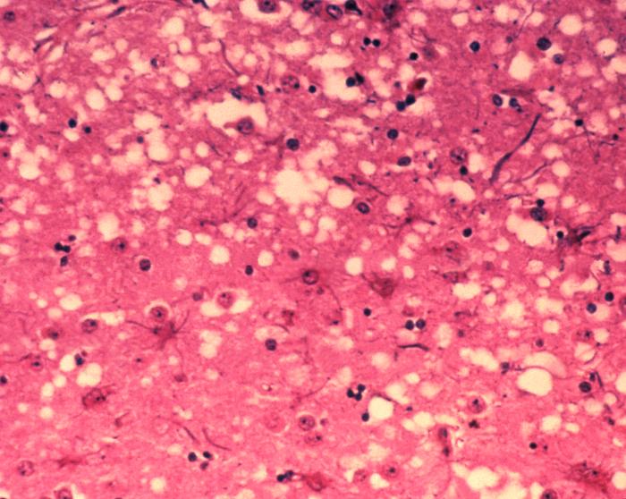
English: This micrograph of brain tissue reveals the cytoarchitectural histopathologic changes found in bovine spongiform encephalopathy. The presence of vacuoles, i.e. microscopic “holes” in the gray matter, gives the brain of BSE-affected cows a sponge-like appearance when tissue sections are examined in the lab.
Nederlands: Deze microscopische opname toont hersenweefsel van een koe die aan BSE gestorven is. Tussen de hersencellen ziet men duidelijk verschillende vacuoles, die deze coupe (weefselsnede) een sponsachtig aanzicht geven.
Deutsch: Das Bild zeigt die histopathologischen Veränderungen die bei einer Infektion mit BSE auftreten. Die Vakuolen, die in der grauen Substanz (substantia grisea) auftreten geben dem Bild ein schwamm-artiges Aussehen.
Français : Cette coupe de tissu cérébral montre les modifications histopathologiques de l'organisation cellulaire lors d'une encéphalopathie spongiforme bovine. la présence de vacuoles, c'est-à-dire des "trous" microscopiques dans le tissu cérébral, donne au cerveau de vaches atteintes de l'ESB un aspect en éponge à l'examen des tissus en laboratoire. |
| 날짜 | |
| 출처 | Public Health Image Library, APHIS: http://www.aphis.usda.gov/lpa/issues/bse/bse_photogallery.html |
| 저자 | Dr. Al Jenny |
| 다른 버전 | http://en.wikipedia.org/wiki/Image:Aphis.usda.gov_BSE_5.jpg |
|
|
 |
파일 역사
날짜/시간 링크를 클릭하면 해당 시간의 파일을 볼 수 있습니다.
| 날짜/시간 | 섬네일 | 크기 | 사용자 | 설명 | |
|---|---|---|---|---|---|
| 현재 | 2005년 6월 21일 (화) 19:21 | | 700 × 558 (73 KB) | wikimediacommons>Obarskyr | {{PD}} |
이 파일을 사용하는 문서
다음 파일 1개가 이 파일과 중복됩니다 (자세한 정보):
이 파일을 사용하는 문서가 없습니다.